Quantcast
Channel: Parasite of the Day
Viewing all 243 articles
Browse latest View live

Sphaerularia vespae

$
0
0
Hornets can put fear into the minds of many people, but today meet the parasite that the hornets fear (if they are capable of fear). Sphaerularia vespae is a parasitic nematode that infects the Japanese yellow hornet (Vespa simillima) and as far as infection goes, this one is quite a doozy. It specifically invade and resides in the gaster (abdomen) of female hornets where it grows and develop. The nematode ends up sterilising the host (much like other parasiticcastrators we have featured on this blog), turning her into a cozy nursery for baby worms. But a new study has shown that they are capable of doing more than just castrate the hornet.

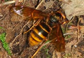
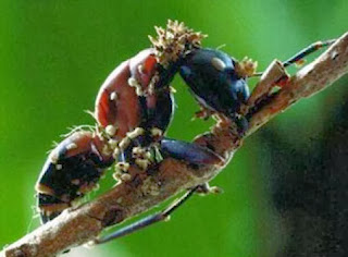
Photo of a queen hornet (from Fig. 2 of the paper)
In a previous study, a group of scientists noticed that the majority of overwintered hornet queens caught in bait traps were infected with S. vespae, so there is something about these nematode-infected hornets which seems to make them more likely to end up in those traps. During autumn/fall, queen hornets fortunate enough not to be infected with S. vespae would visit and poke around various nooks and crannies (usually decayed logs) in the forest to find a spot to hibernate. When the hornet find a place she likes, she will start excavating a hibernacula ( a place to hibernate) and line it with plant fibres that serve as nesting material. But queens that are parasitised and sterilised by S. vespae start visiting decaying logs much earlier during early to mid-summer.

A team of scientists in Japan decided to find out just what those infected queens are up to. For three months between May and August, they made regular weekly visits to a predesignated sites in a forest at the foot of Mount Moiwa and set up a video cameras to observe the decayed logs in the morning and afternoon.

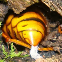
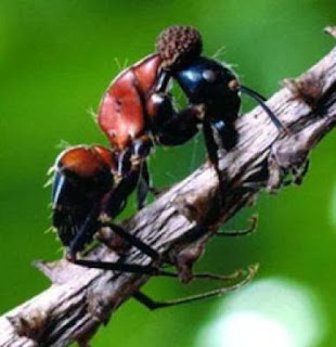
Photo of a hornet releasing
some S. vespae juveniles
(from Fig. 2 of the paper)
They saw that unlike other hornets, the nematode-infected queens never dig nor gather nesting material. They simply crawl inside a decayed log, hang out for a while, then fly off. That is because they have become sterilised couriers that visited potential hibernation sites only to drop off a special package in the form of S. vespae juveniles. A quarter of the infect queens they saw landing on decayed logs offloaded some nematodes (there were some hornets that moved out of sight so the scientist couldn't see what they were up to). But in addition to those observations, the scientists also captured some hornet queens and brought them back to the laboratory for further examination. They kept them in vials and noticed that over two-third of the infected hornets ended up releasing juvenile worms.

When they dissected hornets to see how many of them were infected and to check the developmental stage of their parasites, they found a seasonal pattern to the infections. Queens caught during May and June were mostly infected with fully-mature female worms and their eggs, while queens caught between July and throughout August were filled with juvenile worms that were ready to disembark and infect a new host - which just so happen to be the period when parasitised queens start making regular visits to potential hibernation sites.

So that is S. vespae's game - use the hornet as a mobile incubator/nursery, fly her around during summer to scope out the best pieces of real estate around the forest, then drop off a bundle of worms that can lie in wait like a booby-trap for an uninfected hornet queen to come along and settle in for winter. To complete its life cycle, S. vespae simply take advantage of a preexisting behaviour (seeking out hibernation sites) from the host's repertoire, and "switch it on" at a different time of year to fit the developmental schedule of the parasite's own offspring. Parasite manipulation isn't necessarily about teaching an old host new tricks, but to get the host to perform the tricks that it already knows in a brand new context.

Reference:
Sayama, K., Kosaka, H., & Makino, S. (2013) Release of juvenile nematodes at hibernation sites by overwintered queens of the hornet Vespa simillima. Insectes Sociaux60: 383-388.

Ophiocordyceps sessilis

$
0
0
There are many species of fungi that infect insects and some of the most well-known species are the ones that infect ants, better known to most as the "zombie ant fungus". We have previously featured one such fungus and its ant-jacking antics on this blog. But while most people might think that there's just a single zombie ant fungus out there which is responsible for creating this intriguing wonder (or nightmare) of nature, there are actually many different species of such fungi and they are found all over the world infecting various different insects. In the Ophiocordyceps genus alone there are over a hundred species and there might be some undescribed fungi that are hiding in plain sight because they have been misidentified and misclassified as a previously known species.
Photo of Ophiocordyceps sessilis from
Fig. 1 of the paper

Today, we are going to be featuring one such fungus and it hails from Japan where they are called Kobugata-aritake which means the "bump-neck ant fungus". The fungi specimen described in the paper we are discussing today were originally collected in 2006 from a forest near the village of Iitate, Fukushima. They were initially thought to be specimen of a fairly commonly found species call Ophiocordyceps pulvinata, but upon reexamination, researchers noticed a number of key differences which separated O. sessilis from O. pulvinata.

Both fungi were found sprouting from dead ants which had their mandibles clamped tightly around a branch in the typical "zombie ant" pose, but whereas O. pulvinata produce a bulbous fruiting body that sprouts from the back of the ant's head (see photo on lower left), ants infected with O sessilis are covered in spiny fruiting bodies jutting out all over the ant's body (see photo on upper right).

Further difference between the two fungi can be seen under the microscope; O. pulvinata produce discrete spores that are long and slim, but the spores of O. sessilis look like beads on a necklace which readily breaks apart into small "part-spores". These part-spores of O. sessilis can also germinate on malt-extract agar plates within two days, growing into soft, velvety colonies of fungal mass, whereas O. pulvinata spores failed to grow on such artificial medium. Finally, comparisons of sequences from selected genetic markers revealed that O. sessilis is clearly a very different species to O. pulvinata.

Photo of Ophiocordyceps pulvinata from
Fig. 1 of the paper
A peculiar thing the researchers noticed is that O. sessilis is only ever found in ants that are also infected with O. pulvinata. They suggested that O. sessilis is actually a parasite of O. pulvinata itself and noted other Ophiocordyceps species are often found in pairs, so what had previously be considered as coinfections may in fact be a case of hyperparasitism (whereby a parasite is itself infected by a parasite).

However, there is another possibility that the researchers did not mention in their paper, which was that O. sessilis needs O. pulvinata to pave the way in order for them to colonise the ant's body. An example of this is can be found among fluke-snail host-parasite systems. Like most digenean trematodes, the blood fluke Austrobilharzia terrigalensis they needs to infect a snail for the asexual part of its life cycle, but unlike those other species, A. terrigalensis cannot infect a snail on its own and is always found in snails that are already infected with another species of fluke. The coinfecting species always appear shriveled and emaciated in the presence of A. terrigalensis and it has been suggested that while A. terrigalensis lacks the ability to subvert or suppress the immune defences of snails, they are capable of colonising a snail once its defences have been knocked out by another species, at which point they barge in, overpower the resident parasite and take over the host.

So either O. sessillis is a hyperparasite (or a "mycoparasite" - a parasite of a fungus) of O. pulvinata, or it cannot colonise a host on its own and instead piggybacks on O. pulvinata, eventually usurping it and taking over the ant for its own. Either way, it appears that O. sessilis is a fungus that can hijack a fungus which is used to hijacking ants.

Reference:
Kaitsu, Y., Shimizu, K., Tanaka, E., Shimano, S., Uchiyama, S., Tanaka, C., & Kinjo, N. (2013). Ophiocordyceps sessilis sp. nov., a new species of Ophiocordyceps on Camponotus ants in Japan. Mycological Progress 12: 755-761.

P.S. I recently wrote an article for The Conversation about parasites that can survive freezing - including the hairworm (otherwise known as the parasite that gives crickets nightmares). To read it, just follow this link here.

Tracheliastes polycolpus

$
0
0
Photo of adult T. polycolpus from here
Tracheliastes polycolpus is a parasitic copepod that live on freshwater fish and do so by attaching to the fins of their host, grazing on mucus and epithelial cells. While T. polycolpus can infect handful of different freshwater fishes, it is primarily found on the beaked dace (Leuciscus burdigalensis). When they occur in large numbers, their feeding activities can severely erode the fins of their hosts, so much that in some fish the fins gnawed down to mere nubs (see the photo below of a heavily parasitised dace, with outlines showing the missing fin tissue).

So when it gets crowded on this parasite's usual, preferred host, some T. polycolpus find a home elsewhere and start parasitising other species of fish living in the same area. Even though T. polycolpus is considered to be a host generalist and can infect multiple species of fish, not all fish are considered equally habitable for this parasite and it does have a predilection for certain species over others. So what determines which other fish end up acquiring these parasitic copepods?

A group of scientists from France conducted a study looking at T. polycolpus population on freshwater fish in two French rivers, focusing on the 10 most abundant fish species in those rivers. Of the fish that they examined, eight of them were cyprinids (the family of fish that include dace, roach, and carp) while the two remaining species were the stone loach and brown trout.
Photo of parasitised dace with missing fin tissue from this paper

Only cyrpinids were found to be infected with T. polycolpus and of those only four species (dace, nase, gudgeon, minnows) were found to be consistently infected across both study sites. It turns out that next to the beaked dace, the second most preferred host for T. polycolpus is Parachondrostoma toxostoma, also known as South-west European Nase. After the beaked dace, it was the most commonly infected fish, especially in the Viaur river where there was generally higher abundance of the parasite.

It just so happen that out of all the fishes in those rivers, the nase is most similar to the dace in terms of its general body size, feeding style and habitat, making it the ideal second choice for T. polycolpus. On the flip side, it seems that minnow is the worst host for T. polycolpus - it hosted the least parasites out of the four fish species that were found with T. polycolpus and the parasites that were found on minnows were smaller and produced less eggs than those found on the other fish species. This is probably due to the minnow being a smaller fish than the beaked dace or the nase, so it does not produce as much mucus for T. polycolpus to graze on.

So even when generalist parasites do infect other hosts, they prefer some familiarity. The more similar you are (physiologically and/or ecologically) to the parasite's preferred host, the more likely that you will be next in line to get infected should the parasite's preferred host become too heavily parasitised.

But here's an added to layer to this story which you might want to consider - the South-west European nase is actually listed as a vulnerable species - its population has declined by at least 30% in the past 10 years due to habitat destruction and hybridisation with introduced species, so if the number of nase continues to decline, what does this mean for T. polycolpus? Would this result in increased parasite pressure on other fish species as they find themselves soaking up the "excess"T. polycolpus? Or will the the beaked dace experience even more exacerbated pathology as T. polycolpus are left with less alternative hosts to infect?

Reference:
Lootvoet, A., Blanchet, S., Gevrey, M., Buisson, L., Tudesque, L., & Loot, G. (2013). Patterns and processes of alternative host use in a generalist parasite: insights from a natural host–parasite interaction. Functional Ecology27: 1403-1414

Lethacotyle vera

$
0
0
Images from the paper 
While "many sucker-cups at the rear" sounds like the description for a Lovecraftian monstrosity, that is the name of a group of monogenean parasites called the Polyopisthocotylea. Let's just refer to them as "Poly-Opees" from this point to avoid that tongue-twister. They are ectoparasitic flatworms usually found on the gills of marine fish. Seeing as fish use their gills to extract oxygen from their aquatic environment, there is a constant flow of water washing over these parasites, which means these flatworms are essentially living in a high-flow environment. To secure themselves to the gill filaments, they have a sucker structure on their rear - this sucker anchors the worm in place, allowing it to flex the rest of its body and browse on gill tissue and blood.

The rear suckers of monogeneans are not just a simple suction cup, but are composed of an array of intricate anchors, hooks, and clamps that vary considerably between different groups. In the case of the Poly-Opees, this sucker is armed with a series of clamps that gives that entire group its name. But today we are featuring a species that completely bucks that trend. Like most other Poly-Opees, it is also found on the gills of fish, but stands out due to the complete lack of clamps on its rear sucker.

Lethacotyle vera is closely related to a monogenean that was originally described over sixty years ago. The first species described from the genus Lethacotyle was Lethacotyle fijiensis - which was described from a unspecified carangid fish from Fiji (note to fellow scientists - please take detailed notes!), but there are only four specimens of this parasite in existence and only one of them is stored in a museum available for researchers to examine.

A group of researchers revisiting this species' description noted the unusual absence of clamps on its rear sucker and decided to follow up the lead to look for this mysterious monogenean (or at least a related species - which was what they found). As L. fijiensis was originally described from a carangid fish (the group which include jacks, pompanos, trevally and scad), they decided that's where they should start looking. They obtained some Brassy trevally (Caranax papuensis) from some amateur fishermen and fish markets at New Caledonia and looked through the fish's gills for monogenean parasites.

In was on the gills of those trevally that they came across the new species we are featuring today. They were able to confirm that monogeneans in the Lethacotyle genus do indeed lack clamps compeltely on their rear end. Poly-Opees vary in the number of clamps they have - some species have dozens of well-developed clamps while others have clamps that are rather small and may even be considered as vestigial. In the case of Lethocotyle, they are completely gone.

But if they have no clamps, how do they hang on? They have four tiny hooks on their rear, but they are so small that they probably contribute little to securing the worm in place. The researchers noted that instead, the rear sucker has turned into a flap covered in "tegumental striations" in the place of clamps. These are microscopic wrinkles that increase friction and provide traction against a substrate - these microscopic structures might be somewhat comparable to those found on the foot pads of some insects. In this case, it provides enough traction to keep L. vera securely fastened to the gills of its host.

What the story of the Lethocotyle genus and their rear suckers shows us is that parasites are far from being "simplified" evolutionary dead ends, but that they continue to evolve new structures even as they shed others. As with free-living species, certain features often become lost or vestigial over the course of evolution, but then new structures evolve in their place. Lethacotyle might have lost its clamps, but it has also gained a new attachment feature (striation-covered flap) that makes it unique among all the known monogeneans.

Reference:
Justine, J. L., Rahmouni, C., Gey, D., Schoelinck, C., & Hoberg, E. P. (2013). The Monogenean Which Lost Its Clamps. PloS one, 8(11): e79155.

Another year of parasites in insects, in shellfish and in extreme environments

$
0
0
It is hard to believe that it's already been another year again, and it was a particularly exciting year too, with a lot happening with and around this blog. In terms of the parasites we featured on here, there were some which can be considered to be pretty extreme; like the only external parasite found on guppies that live in noxious tar pits, and some tapeworms with an special affinity for heavy metal. There are those that might make your squirm; like the sexually-transmitted roundworm in anole lizards, and a crustacean that lives in a fish's bladder.

We gave seafood fans some food for thought with some parasites that plague catfish and flounder, and checked in on bunch of clam parasites (tapeworms and flukes) and mussel parasites too (Himasthla elongata). And while fish and shellfish might provide some fodder for parasites, on land, insects provide plenty more opportunities for parasitism, after all, insects are the most diverse group of animals on Earth and they make abundant hosts; from crickets to hornets to ants, and amongst these parasite of insects (some of which are insects themselves) there are some rather sinister ones - like the parasitoid wasp that takes its host to the edge of death so it can be a more compliant host, or the mosquito-killing round worms which sit like mines to be activated upon detecting the presence of its mosquito larva host.

Of course, this year we also had some guest bloggers in the form of students from the University of New England ZOOL329/529 class of 2013 who wrote about how toxic birds makes for sad lice, self-medicating in bees, avian malaria parasites that make their host more attractive to mosquitoes, and how an intertidal fluke might respond to a rise in global temperature. Also, as with last year, we brought you some conference coverage too (part 1, part 2).

We will be back next year with plenty more posts about the newest research in fields relating to parasitology which you might not have heard or read about elsewhere, and as usual, I have already lined up a few which I am going to be writing about... See you all next year!

P.S. If you can't wait until next year, you can find some of my other parasite-related writing on The Conversation about freeze-tolerant parasites, a worm that usurp hornet queens, and fungi that plague the zombie ant fungus. And alongside writing this blog, I've doing a regular radio segment call "Creepy but Curious" where I sometimes talk about parasitic (among other things), like the zombie ants, the infamous crab-castrating Sacculina, the tongue-biter parasite, and the virus that melts caterpillars.

Choniomyzon inflatus

$
0
0
Photo of C. inflatus from the paper
I guess you can kind of say the parasite we are featuring today is a "balloon animal" and indeed its name refers to it. According to the paper which described and named this copepod - Choniomyzon inflatus - "The specific name of the new species is a reference to its swollen prosome, which resembles a balloon."

But you won't be finding this odd little crustacean at any kid's party, instead it is usually attached to the egg masses of the smooth fan lobsters (Ibacus novemdentatus) on the coast of western Japan. It is the third species from the Choniomyzon genus to have ever been described and there are two other known species; C. panuliri which are found on spiny lobsters from India, the British Solomon Islands and the Great Barrier Reef, and C. libiniae which lives on spider crabs from São Sebastião Island, Brazil. All three species live by attaching themselves to the external eggs masses of their respective hosts.

SEM photo of C.inflatus
from the paper
So why do they look like a miniature hopper ball toy? Well, that relates to where they live and what they feed on. Chioniomyzon inflatus belongs to a family of copepod called the Nicothoidae and the reason they do this Humpty Dumpty impersonation is so that they can insinuate themselves amidst the eggs masses of larger crustaceans.

Normally the host crustaceans would remove any foreign particles or organisms that get caught up in their brood pouch or egg mass, but by disguising itself as an egg, C. inflatus and its relatives can stay there undisturbed. And while its appearance seems comical to us, it is seriously bad news for its host because nicothoid copepods are egg-eaters - they have a syringe-like mouthpart with which they puncture their host's eggs and suck out their contents.

So C. inflatus masquerades as just another egg in the brood to avoid being expel meanwhile munching on the actual eggs around it. This strategy is rather reminiscent of another creature that we featured during the first year of the Parasite of the Day blog - the cuckoo catfish which hides its eggs amongst that of mouth-brooding cichlids. You can read more about the cuckoo catfish here.

Reference:
Wakabayashi, K., Otake, S., Tanaka, Y., & Nagasawa, K. (2013). Choniomyzon inflatus n. sp.(Crustacea: Copepoda: Nicothoidae) associated with Ibacus novemdentatus (Crustacea: Decapoda: Scyllaridae) from Japanese waters. Systematic parasitology 84: 157-165.

Phronima sp.

$
0
0
Today's guest post is by Katie O'Dwyer, a PhD student currently at University of Otago in the Evolutionary and Ecological Parasitology research group. In one of my conference reports last year, I mentioned some of the research that she is currently conducting on parasitic flukes that live in periwinkles. She has provided us with a post about a parasite that she came across while walking along a beach in New Zealand.

Phronima and its salp barrel.
Photo by Katie O'Dwyer, used here with permission
After recently finding some salps containing the amphipod Phronima, washed up on a beach in New Zealand, I decided this was a worthy group to compose a blog about. It helped too that I was already interested in this group of crustaceans, having assisted with some work on them in Ireland. Read on for some interesting information on this little studied group of parasitic organisms…

Imagine a parasite which can create its own mobile nursery for its young, a parasite which is thought to be the inspiration behind the chestbusting xenomorph in the popular movie Alien. Well imagine no more! Introducing Phronima, the pram bug. These amphipods are members of the Phronimidae, a group of ten species of hyperiid amphipod, which occur in the water column throughout the open ocean. This sets them apart from their close relatives, which typically inhabit the benthic environment of the seafloor. So what has allowed this particular family to adapt to the pelagic or open water environment?

Those adorable little babies!
Photo by Katie O'Dwyer, used here with permission
Enter salps. What is a salp? Salps are gelatinous zooplankton which drift throughout our oceans. They may occur singly or in huge chains composed of individual salps linked together. Phronima is equipped with impressive front claws and with these they attach to an individual salp and carve away its insides until it forms a barrel. Phronima then climbs inside and sails the sea from inside a gelatinous barrel, collecting food from the water column. A number of questions may now come to mind regarding this symbiosis; has Phronima killed its host, which suggests that it is a parasitoid rather than a parasite, and why does it carry this barrel around as it must be pretty energetically expensive, right?

Well, as mentioned, these organisms live in the open ocean which presents several challenges to collecting samples for answering these questions. However, some dedicated researchers have indeed managed to study these fascinating creatures on the rare occasion that such an opportunity arises. From their research they have found that the salp in fact still contains live cells, although it hardly resembles a salp anymore with just a barrel of tissue remaining. The presence of live cells means that the barrel maintains its structure and that is important for Phronima to have a sturdy home. As the barrel barely resembles a live salp any longer, Phronima should really be considered as parasitoids rather than parasites.
Do a barrel roll!
Photo by Katie O'Dwyer, used here with permission

As for the energy involved in carrying around this barrel, the barrel provides a larger structure than the amphipod itself and this enables the Phronima to be more buoyant in the water column. However, some energy is still required to carry around this jelly barrel. Overall energy usage by Phronima is higher than that of benthic amphipods but on the lower spectrum compared with other pelagic or open water amphipods. This suggests that Phronima have indeed adapted to a unique niche which enables them to travel in the water column with their young and access new food resources without this behaviour being too energetically costly.

One unusual finding in the research thus far is that male Phronima are also found in barrels. If Phronima is known as the pram bug, which suggests the barrel is important for carrying offspring, then why should males carry a barrel too? Could they use it as part of some mating strategy, where they pass the barrel on to the female they mate with? Due to the difficulties associated with studying organisms that dwell in the open ocean many questions remain unanswered and this leaves us ever more curious and fascinated by creatures such as Phronima.

References:
Hirose, E., Aoki, M. N., & Nishikawa, J. (2005). Still alive? Fine structure of the barrels made by Phronima (Crustacea: Amphipoda). Journal of the Marine Biological Association of the United Kingdom 85: 1435-1439.

Bishop, R. E., & Geiger, S. P. (2006). Phronima energetics: is there a bonus to the barrel? Crustaceana 79: 1059-1070.

This post was written by Katie O'Dwyer.

Daubaylia potomaca

$
0
0
Photo is of a related species,
Daubaylia malayanum from here
For a parasite, the host provides provides food, shelter, and a site for reproduction - in short, a complete habitat. While for some parasites, host death is a necessary condition for the parasite to complete its life-cycle, for others, the death of a host amounts to the end of the world (or a sinking ship at the very least).

Meet Daubaylia potomaca, a roundworm which infects the freshwater snail Helisoma anceps. Unlike other roundworms that use snails as vehicles to reach the next host in their life-cycle, the snail is the only hosts for D. potomaca. But seeing as snails do not live forever, any parasite it harbours would need an exit strategy or risk perishing with their host when the end finally comes. For a parasite like D. potomaca which completes its entire life-cycle in the snail, it would be useful for it to recognise when they should abandon their host.

A dying host is not the only reason to leave - finding a new host is integral to most parasites' life-cycle, but you would not want to leave too early either - the outside world is a hostile place and as a parasite, you would want to get as much out of the host as possible before you make a run for it. Unlike most other parasites that usually infect a new host as larval stages, D. potomaca actually leave their hosts as fully-matured females laden with eggs, all tangled in mucus-coated bundles composed of 10-50 worms. Therefore the female worms would not want to depart too early as it needs to gather as much resources as possible from its host to nurture the developing eggs. So ideally, they leave it to the last possible moment before they emerge from the snail.

So how well does D. potomaca time its escape? In the paper we are featuring today, a team of researchers studying this host-parasite system observed some worms leaving as early as 52 days before their host snail died, but the majority (almost a quarter) of the worms came out in the last five days of the snail's life. The percentage of worms that emerged increased as the snail's life draws closer to its end - it seems almost as if the parasite can sense when the host is near death's door and took that as a cue for when to bail.

As an additional factor, the researchers also found that infection intensity of D. potomaca affected the snail's lifespan - the more heavily infected it is, the sooner the snail dies. So perhaps D. potomaca can also gauge how crowded the inside of the host is, and schedule their departure accordingly. This is some what reminiscent of a parasite previously featured on this blog; Coitocaecum parvum.

Unlike D. potomacaC. parvum is a fluke with a complex life-cycle and infects multiple hosts through out its life. However, it does face the potential problem of its amphipod host dying before it is eaten by the parasite's next host, in this case a freshwater fish where it can mate with other flukes and produce eggs. But if become increasingly unlikely that its amphipod host would be eaten by a fish before it expires, C. parvum would alters its usual life history schedule to start producing eggs in the amphipod instead of waiting until it end up in the gut of a fish (which might not happen).

While it is a different kind of response to imminent host death compared with D. potomaca, it is another example of how parasites can assess the status of its host and the surrounding environment, and adjust their own life schedule accordingly. Throughout the course of co-evolving with their hosts, in addition to adapting to their host's defences, parasites have also developed many strategies to ensure their survival even as their environment (i.e. the host) faces imminent collapse.

Reference:
Zimmermann, M. R., Luth, K. E., & Esch, G. W. (2013). Shedding Patterns of Daubaylia potomaca (Nematoda: Rhabditida). Journal of Parasitology 99: 966-969.

Gordionus chinensis

$
0
0
Hairworms are known for their ability to make their host go for an impromptu (and terminal) swim in a stream or a pond, but by doing that they are not just sending ripples through the water, but also into the surrounding ecosystem. The paper we are looking at today features a species of hairworm from Japan call Gordionus chinensis - this parasite infects three different species of forest-dwelling camel crickets from the genus Diestrammena.

Photo by Danue Sachiko from here
The scientists who conducted the study that this paper is based on wanted to find out what happens to the the cricket population and their hairworm parasites after their home forest has been cut down. They conducted an observational field study at an experimental forest in the upper parts of the Totsu River at Nara Prefecture, Japan. The forest was originally clear-cut in 1912 and 1916 and since then, parts of it have been replanted and cut down at different point in time over the last century. Each study site corresponds with a different replanted forests of Japanese cypress ranging from 3 to 48 years old.

These scientists found that the camel crickets began returning a few years after a forest has been replanted, their abundance steadily increasing and eventually reaching a peak after the forest has been standing for at least 30 years. But their hairworm parasites did not return with similar gusto. In fact, they estimated that only second-growth forests that are more than 50 years old have hairworm populations that are as abundance as those found at undisturbed sites.

One possible reason for the hairworms' slow recovery is their complex life cycle which requires infection of more than one host. The replanted forest might be lacking some of the other host G. chinensis needs to complete its life cycle. Because parasites has such a negative public image, a forest which is free of parasites (or at least a specific parasite) might sound good to most people. But these hairworms actually play a very vital role in the ecosystem.

By causing their cricket host to jump into a stream, they actually serve as a kind of fast food delivery service for the fish living in those streams. A cricket infected with a hair worm is 20 times more likely to stumble into a stream and become fish food than an uninfected cricket - so fish which would not usually get to feed on such large land-loving insects on a regular basis can now do so thanks to the hairworm, and it has calculated that this straight-to-your-stream food delivery service accounts for 60% of the trout population's energy intake in some watersheds.

For hairworms, new forests just do not have the same creature comforts of old forests. And if you are a keen angler or simply appreciate a fish-rich stream - you have a parasite to thank for all the fishes.

Reference:
Sato, T., Watanabe, K., Fukushima, K., & Tokuchi, N. (2014). Parasites and forest chronosequence: Long-term recovery of nematomorph parasites after clear-cut logging. Forest Ecology and Management, 314: 166-171.

Cucumispora dikerogammari

$
0
0
Invasive species can be very disruptive - cane toads, rabbits, water hyacinth, and zebra mussels are just a few well-known examples of species that have been introduced to areas outside of their original geographic range and have caused extensive ecological disruption in their new home. One of the hypotheses for why some introduced species become so successful when they arrive at a new region is called the "enemy release hypothesis". In their new home, introduced species run amok as they are no longer hounded by their usual foes that would otherwise keep their population in check.
Top: A heavily infected amphipod
Bottom: Spores of C. dikerogammari
Photo from here

Dikerogammarus villosus is an amphipod (a little, shrimp-like crustacean) from the Ponto-Caspian that has invaded western and central Europe, and is now also found in the United Kingdom. They might only grow up to a little over an inch long, but they are voracious little predators that eat everything smaller than themselves, including each other. Released from their usual predators and parasites, D. villosus rips through the freshwater life of its new neighbourhood. But they have not completely escaped from their past foes; one parasite has managed to come along for the ride, and it is a microsporidian called Cucumispora dikerogammari.

As far as the parasite goes, Cucumispora dikerogammari is a pretty nasty one. It invades the host's muscles, reproduces prolifically and eventually kills the host by overwhelming it with sheer numbers. There is some concern that this parasite can spill over into the native invertebrates and add insult to injury to the local stream life. But on another hand, a new study shows that this parasite might be one of the few things holding back this voracious invasive amphipod from causing even more destruction.

A group of scientists from France conducted a study to looked at how C. dikerogammari affects the activity levels and appetite of D. villosus. They observed the behaviour of both infected and uninfected amhipods in a water-filled glass tube and noticed that amphipods at a late stage of infection that are visibly "filled to the brim" with parasite spores are actually more active than healthy amphipods or those that are not visibly parasitised because they are at a much earlier stage of the infection.

Close-up of a C. dikerogammari spore from here
Furthermore, they also presented amphipods with midge larvae (also known to some as "bloodworms") to see how many they ate. Both infected and uninfected D. villosus pounced on those insect larvae, but the heavily infected amphipods ate far less than the healthy ones. For whatever reason, this parasite seems to cause D. villosus to lose its appetite, and given this crustacean's reputation of eating everything that it can get its claws around, this may have significant ecological ramifications. It could mean that C. dikerogammari may be subtly reducing the impact these amphipods have on the areas where they have been introduced.

But why would heavily-infected D. villosus, which would have much of their muscle mass already converted to parasite spores by C. dikerogammari, be more active? Well, it could just be an odd manifestation of the disease, but if it is, it is certainly a useful one for this parasite - as it depends upon cannibalism for transmission to new hosts. Dikerogammarus villosus are rather homely creatures and usually prefer to stay under a shelter and wait for potential prey to wander by. By getting their host out and about, C. dikerogammari might increase the chances that its host will either run into one of its cannibalistic buddies, or die out in the open where it can be scavenged by other D. villosus.

It seems that for this little invasive amphipod, no matter how far you go, you can never really run away from your past (foes).

Reference:
Bacela-Spychalska, K., Rigaud, T., & Wattier, R. A. (2013). A co-invasive microsporidian parasite that reduces the predatory behaviour of its host Dikerogammarus villosus (Crustacea, Amphipoda). Parasitology141: 254-258.

Octopicola superba

$
0
0
When it comes to reproduction, most living things can be classified along a scale. At one end, you have the r-strategists (many insects and molluscs) that produce a prodigious number of offspring but few survive to adulthood. And on the other end are the K-strategists that produces only a few progeny, but to invest a lot of resources into each to ensure they are more likely to reach maturity (for example, elephants, humans, etc).

SEM photo of female
Octopicola superba from here
There is a cost/benefit trade-off inherent with being on either side of the scale because as a r-strategist, you might be producing a lot of progeny, but most of them will probably die before they get to reproduce themselves. While on the K-strategist end, by investing so much resources into each individual young, you can only afford to produce a few of them. The reproductive strategy of different organisms all fall somewhere along that continuum between low quality mass production or high quality but infrequent output, and different circumstances call for different strategies.

Textbook often use parasites as key examples of r-strategists, as a model of organisms that producing prodigious number of offspring. Indeed some internal parasites are well-known for their reproductive capacity - for example, the female blood fluke Schistosoma mansoni produces 300 to 3000 eggs per day, while tapeworms like Diphyllobothrium dendriticum can produce tens of millions of eggs per day. But not all parasites opt for quantity over quality.

The study we are featuring today examined the reproductive capacity of the parasitic copepod Octopicola superba, which, as its name indicates, lives in the common octopus. As far as a parasite goes, this crustacean seems rather innocuous and does not really cause much harm to its host. Octopicola superba can be found all over the body of the octopus but most of them are located on the skin and gills. Even though it is a parasite, it has a reproductive strategy which brings it closer to being a K-strategist.

Each female O. superba produces a clutch of only a few dozen eggs per season; if a female was to produce more than about forty eggs in a clutch, she starts reaching the upper limit of her reproductive capacity and the size of each egg (which reflects how much resources is invested into it) begins to shrunk as the brood imposes too much of a drain. This reproductive capacity varies considerably between individual; the most productive copepods are able to produce over twice as many eggs as the least productive ones, while some produced eggs that were almost twice as big as those produced by others.

Octopicola superba's reproductive strategy also shifts during different seasons; in winter, they produced a larger clutch of smaller eggs, whereas in summer they produce a smaller clutch of bigger eggs. Such season shifts has been observed in other parasitic copepods, though for O. superba, the reason for them doing so remains unknown. Despite these seasonal and individual differences, overall O. superba is certainly low-key when it comes to reproduction - even the most fecund female had just above sixty eggs in a clutch and the rest mostly produced between thirty to forty eggs.

So why has this parasitic copepod evolved to produce so few eggs compared with parasites like tapeworms and blood flukes that pump out thousands or even millions of eggs on a daily basis? It might have something to do with the habits of its host.

Octopus tend to be territorial homebodies that likes to stay in their little corner of the sea. Previous analyses indicate that hosts with such sedentary habits tend to select for parasitic copepods that produce larger eggs. Unlike one infecting more mobile animal (like a fish), parasites of sedentary animals cannot rely upon their host's routine daily movement to bring them into contact with new hosts. Therefore, they must do so under their own steam. By investing more into each egg, the female O.superba ensures each of her babies are better equipped for the long journey to find a new home, even if it means she can only produce just a few dozen of them at a time.

With offspring, you can only invest so much into them - at some point, they are on their own

Cavaleiro, F. I., & Santos, M. J. (2014). Egg number-egg size: an important trade-off in parasite life history strategies. International Journal for Parasitology44:173-182

Bivitellobilharzia nairi

$
0
0
A little over a year ago, I wrote a post about Bivitellobilharzia loxodontae - a species of blood fluke that lives in the African forest elephant. Today I am writing about a study on another species from that genus - Bivitellobilharzia nairi - which parasitises the Indian elephant. However in a newly published study, it turns out the Indian elephant is not the only thick-skinned mammal that harbours this fluke.

Photo of Indian rhino by Krish Dulal
The study we are featuring today took place in southern Nepal at the Chitwan National Park (CNP). Researchers collected fecal samples from both domesticated and wild Indian elephants for examination and as expected, they found B. nairi eggs amongst the samples. But it was when they started looking in the poop of Indian rhinoceros that they found the unexpected. These rhinoceros do not take a dump just anywhere; they are creatures of habit and defecate at specific spots call faecal middens - which is how they mark their territory. When the researchers dug through the contents of those middens, they found blood fluke eggs amidst the rhino dung in half of the fourteen middens they sampled from.

The eggs had the characteristic look of schistosome eggs - an ovoid with a hook at one end (see below). But they were not just any blood fluke eggs, they looked very similar to the eggs of B. nairi - the elephant blood fluke. When the researchers sequenced specific marker section of the fluke eggs' DNA, they found that it matched the known sequences for B. nairi, showing that what is usually thought of as just an elephant parasite can also find a home in the Indian rhinoceros. Furthermore, the B. nairi eggs they recovered from the rhino dung were completely viable, showing that the rhino is a natural and commonly used host for this parasite and that they did not end up there by accident

Image of Bivitellobilharzia nairi egg from here
Evolutionarily speaking, elephants and rhinoceros are fairly far apart on the mammalian tree - the last common ancestor they shared lived about 100 million years ago in the era of non-avian dinosaurs. So what is an elephant schistosome doing in a rhino? Despite their specialised adaptations for living in the circulatory system and evading the immune reactions of their particular hosts, throughout their evolutionary history, schistosome have made a number of leaps across divergent animal taxa. One such jumps had allowed the ancestors of schistosomes to evolve from a sea turtle-infecting parasite into one which live in the blood of warm-blooded animals like birds and mammals. While elephants and rhinoceros have had disparate evolutionary paths for at least a hundred million years, clearly their physiology are similar enough for B. nairi to successfully survive in both. In addition, their shared habitat provided the fluke with plenty of opportunity to encounter and adapt to the rhinoceros.

So there is more than one way for two (or more) different species to end up with the same parasite. You can either share a recent common ancestry, or you can share the same habitat which gives the parasite ample opportunities to cross the evolutionary gulf between different hosts.

Reference:
Devkota, R., Brant, S.V., Thapa, A. & Loker, E.S. (2014) Sharing schistosomes: the elephant schistosome Bivitellobilharzia nairi also infects the greater one-horned rhinoceros (Rhinoceros unicornis) in Chitwan National Park, Nepal. Journal of Helminthology88: 32–40

Controrchis sp.

$
0
0
Extreme weather events can cause significant changes to ecosystems and their inhabitants. When Hurricane Iris made landfall at Belize, it caused widespread devastation in its wake. The study we are featuring today was a part of a larger project to look at how Hurricane Iris affected a population of black howler monkeys (Alouatta pigra) which has been monitored since 1998. In the aftermath of the hurricane, the number of monkeys in the forest decreased by 78 percent until their population began to stablise and increase three and a half years later. But aside from such outwardly visible impacts, there were also other changes afoot within the monkeys themselves.
Photo of black howler monkey by Ian Morton

A team of scientists interested in monitoring the recovery carried out a study to see how this has affected the monkeys' parasites. It is possible that these primates are harbouring higher parasite loads than they did before the hurricane due to the stress of living in a disturbed habitat. The scientists collected samples of monkey fece over the course of 3 years and look for parasite eggs. They also measured the level of cortisol, a hormone associated with stress, present in the fecal sample, and collected data on other aspects of the monkey's behaviour to see if they were associated with their parasite burden.

Photo of Controrchis eggs
from here
The black howler monkeys were found to have five species of roundworms and a species of digenean fluke (based on the presence of their eggs in the monkey poop), but the prevalence and abundance of those parasites were not associated with the level of stress hormones. Instead, nematode (roundworms) prevalence was heavily dependent on population density and the size of the groups in which the monkeys gathered. This is to be expected as these worms are transmitted via accidental ingestion of eggs or larvae from the host's feces. The more monkeys there are around in a given area, the more opportunities for these particular parasites to be passed on. This is similar to what has previously found in other studies on primate parasites. But the only factor that successfully predicted the occurrence of the digenean trematode fluke Controrchis was the amount of leaves the monkeys consumed.

While black howler monkeys usually prefer a diet filled with fruit, in the aftermath of Hurricane Iris there were no fruit-bearing plants in the forest for 18 months. So the monkeys were forced to go on a leaf-based diet instead of the fruit-based one they enjoyed before the hurricane, and the plant most readily available and palatable to the monkeys was Cecropia. These fast-growing leafy plants usually happens to be the first on the scene in the wake of such habitat disturbances. They do not contain as much fibre as other plants and have little in the way of noxious defensive chemicals - which makes Cecropia excellent fodder for the black howler monkeys. Cecropia also contains a lot of what these monkeys need in a balanced diet, so in the absence of fruits, the howler monkeys munched readily on these nutritious greens

But why is the consumption of Cecropia associated with the prevalence of Controrchis? The fluke does not use leaves and vegetation as a mean of transmission (unlike Fasciola the liver fluke), instead, Controrchis uses ants as a go-between to get in their vertebrate host. But these monkeys don't really have a taste for ants, so why is Controrchis prevalence linked to the amount of leaves they have consumed? That is because Cecropia also happens to be myrmecophtyes, or ant-plants. Monkeys that are chowing down those leafy greens are also inadvertently swallowing a lot of ants, which means taking onboard a lot of Controrchis waiting to make a connection with a suitable monkey host.

For another more detailed take on this paper, from the lead author herself, see this post here

Reference:
Behie, A. M., Kutz, S., & Pavelka, M. S. (2014). Cascading Effects of Climate Change: Do Hurricane‐damaged Forests Increase Risk of Exposure to Parasites?. Biotropica 46: 25-31.

Nematocenator marisprofundi

$
0
0
Parasitism is the most common mode of life on Earth and it can found everywhere, in all kinds of environments. Even in extreme places such deep sea hydrothermal vents, amidst hellish geysers pouring out hot sulfide or seeping methane, parasitism carries on as usual - the players may change, but the game stays the same. While on this blog most of the nematodes we have featured are the parasites, in this particular case, they play the role of the host.

SEM photo of D. marci from the paper
Laying about 85 kilometres off the coast of Oregon, under about half a mile (800 metres) of water is the Hydrate Ridge methane seeps. These vents are covered in mats of sulfide oxidizing bacteria which are crawling with worms - mostly nematodes from the genus Desmodora. One of these species happens to be a host to the parasite we are featuring today - Nematocenator marsiprofundi - which translates into "nematode eater of the deep sea". It is a microsporidian - a group of single-cell parasite somewhat related to fungi, and taxonomically speaking, N. marsiprofundi lies right near the base of the split between these two groups.

Microsporidians are found in a wide range of animals including vertebrates such as fish and reptiles, as well as invertebrates such as insect, crustaceans, and nematode worms. The host of N. marsiprofundi, a nematode named Desmodora marci (see above), is one of the more abundant animal at methane seeps. There can be as many as twenty worms for each millilitre of carbonate rocks from such locales, and over half of those worms would be infected with N. marsiprofundi. This parasite seems to be common at such vents and were found at sites which are 15 kilometres, so N. marsiprofundi is not localised to just a particular location and/or worm population.

Spores of N. marisprofundi
(image from the paper)
The spores of this parasite (see left) are mostly found in the worm's reproductive tract; in female worms, the spores sit in the uterus next to the eggs, and in the male, the spores lined the worm's sperm duct and cloaca. This led the researchers who found this parasite to suggest that N. marisprofundi is sexually transmitted between its host. However, they also noticed some stages of the parasite were situated in the body wall, where they seem to degrade and digest the worm's muscle tissue, not unlike the microsporidian we featured a two months ago which infects an amphipod that has become invasive in Central Europe.

While their presence in the body wall and the effects they have on their host's muscle indicates they can be quite harmful and may transmit through means other than the worms' sexual activities, that is not to say that this parasite might not exploit multiple mode of transmission. Some parasite change their shape and infect different host tissue at different stages of their lives, and it is possible that N. marisprofundi can both be sexually transmitted and also eventually kill their host to allow their spores to disperse from a rotting cadaver

Studies like this shows parasites might be more common in the deep sea that we might have previously suspected, and that even in seemingly extreme environments like hydrothermal vents, there is good living to had as a parasite. Parasitism is everywhere on this planet, and while many people may think parasites are odd freaks of nature, in reality they are just a normal part of life on Earth.

Reference:
Sapir, A., Dillman, A. R., Connon, S. A., Grupe, B. M., Ingels, J., Mundo-Ocampo, M., Levin, L. A., Baldwin, J. G., Orphan, V. J. & Sternberg, P. W. (2014). Microsporidia-nematode associations in methane seeps reveal basal fungal parasitism in the deep sea. Frontiers in Microbiology 5: 43.

Loxothylacus panopei

$
0
0
Photo by Inken Kruse via the Hare Lab
Some parasites can manipulate their host's behaviour in very spectacular ways, but there are also other parasites that change their host's habits in more subtle manners. While such alteration to the host can seem fairly minor, they can still result in some very profound impact on the rest of the ecosystem.

There is a group of parasitic barnacles call Rhizocephala (the most well-known species is Sacculina carcini) that are capable of castrating their host, turning them into unwitting babysitters that nurture the parasites' brood. The infected crab display some very obvious changes to their behaviour, and in some cases, their appearance. But the study we are featuring today shows that apart from turning them into doting mothers for the parasite's babies, these barnacles can also alter the crab's behaviour in less obvious ways that have ramifications for other marine inhabitants.

The flatback mud crab (Eurypanopeus depressus) lives in estuaries on the coast of South Carolina and it is infected by a species of rhizocephalan call Loxothylacus panopei. In addition to doing the usual host castrating and commandeering trick, L. panopei also changes how this crab responds to potential prey. Usually, the mud crab has an omnivorous diet, dining on algae as well as worms, smaller crustaceans, and sponges. Sometimes they may also have a crack at more armoured prey like mussels. But crabs that are infected with L. panopei lose their appetite for such shell-covered fares.

When researchers offered uninfected crabs with piles of mussels, the crabs acted like they were at an all-you-can-eat seafood buffet and ate as much as they can - the more mussels the researchers presented them with, the more they ate. But no matter how many mussels they offered to crabs that were infected with L. panopei, they simply eat one and call it a day. The parasitised crabs also took longer to get their act together and this seems to be related to the size of the crab's parasite - the larger the parasite has grown, the longer the crab takes to start digging into a mussel.

Based on a field survey of the estuary where the study took place, the researcher concluded that about a fifth of the crab at that location were infected with L. panopei. Given the effects that L. panopei has on their crab's appetite for shellfish, it seems that the mussels might have an unlikely ally in the form a parasitic barnacle. The finding of this study share some parallel to another paper that we featured on this blog earlier this year, on the muscle-wasting parasite that infects a predatory shrimp and curb its otherwise ravenous appetite.

Ecosystems are made up of complicated networks of biological interactions and parasites can mediate predator-prey interactions in different, and sometimes conflicting ways. While some parasites can make prey animals more vulnerable or accessible to predators, there are other like L. panopei that may be reducing the appetite of the said predators. The subtle interplay of such parasite-mediated interactions are often overlooked or ignored, but their effects on the ecosystem are certainly there if you know what to look for.

Reference:
Toscano, B. J., Newsome, B., & Griffen, B. D. (2014). Parasite modification of predator functional response. Oecologia 175: 345-352.

Anilocra nemipteri

$
0
0
Photo from Figure 1 of the paper
The parasite in the study being featured today makes a living riding around on the top of a fish's head and occasionally gnawing on its face. It is in the same family as the infamous tongue-biter, the Cymothoidae, though technically this one is more of a face-hugger.

Anilocra nemipteri is found on the Great Barrier Reef of Australia and it makes a living by hitching a ride (and feeds) from the bridled monocle bream, Scolopsis bilineata. It is a pretty common parasite - in some areas, up to 30 percent of monocle bream carry one of these crustaceans on their head like a nasty blood-sucking beret that stay attached for years.

As you can see from the photo, A. nemipteri is a fairly big parasites comparing with the size of the fish (in some case they can reach as almost one-third the length of the host fish!), and having a parasite of that size hanging off your face is going to be quite a drag - literally. That is bad news for a little fish like the monocle bream that needs to make a quick getaway from any hungry predators on the reef. So just how much of a drag is A. nemipteri? A related species - Anilocra apogonae - which clings to the cardinal fish (Cheilodipterus quinquelineatus) is known to cause their host to swim slower and have lower endurance. Does the same apply for A. nemipteri and the monocle bream?

To find out, scientists compared how quickly the fish can respond to an attack and their Flight Initiation Distance (FID) in both a laboratory setting and in the field. The FID is the distance from a predator at which an animal decides to flee - risk-takers have a shorter FID. They divided the monocle bream into three different groups: parasite-free fish, fish carrying an A. nemipteri, infected fish which just had their parasite removed.

Photo from Figure 1 of the paper
The research team simulated an attack by a bird (with a weighted PVC pipe) on fish in specially-designed experimental tanks and filmed the response to measure the fish's reaction time to the attack. Even though one would think all that face-gnawing from A. nemipteri would have weakened their host, and not to mention the body of the parasite itself causing significant drag, the escape performance of parasitised fish was not all that different from unparasitised - they reacted and got away from the attack just as quickly as their unburdened buddies. In the field experiment, the scientists donned snorkelling gear and tried to approach any monocle breams they spotted and measured how close they could get to the fish before they fled. There, they found parasitised fish have a slightly shorter FID than parasite-free fish, but not significantly so.

Fish that are infected by A. nemipteri are smaller than uninfected ones, and it just so happen that smaller fish tend to allow predators to get closer to them before fleeing. But whether this is due to the parasite is another matter. Are parasitised fish smaller because their growth have been stunt by A. nemipteri? Or does this face-hugger simply prefer smaller fish because larger and older fish might have built up an immunity to it?

Though it may seem less exciting when we find a parasite doesn't cause much behavioural changes in its host, it is vital to our understanding of host-parasite relationships. Perhaps it means the host is able to compensate for the presence of the parasite. Also it is not clear what the long term cost of having A. nemipteri might be over the life time of the fish. It is also important to treat such a case in its context. Unlike other parasite which have a life-cycle and depend upon its host getting eaten by a predator to reach maturity, A. nemipteri is an external parasite that simply sticks to a host and stay for life - if the parasitised fish is eaten by a predator, it'll go down with the host like a bit of garnish and be digested too.

So it is probably just as well that A. nemipteri is not too much of a drag to have around.

Reference:
Binning, S. A., Barnes, J. I., Davies, J. N., Backwell, P. R., Keogh, J. S., & Roche, D. G. (2014). Ectoparasites modify escape behaviour, but not performance, in a coral reef fish. Animal Behaviour 93: 1-7.

Ismaila belciki

$
0
0
Photo of infected Janolus fuscus
used with permission from Jeff Goddard
If you ever find yourself down by the sea, you may come across some very flamboyant sea slugs call nudibranchs. But beneath their colourful exterior, some of them are harbouring a dark secret in the form of a very strange looking parasite. These parasites live hidden inside the main body cavity of their molluscan host, so if you are unfamiliar with this particular critter, you might not even notice it. The main thing that gives away their presence are a pair of egg sacs poking out of the sea slug (see photo on the right). Those egg sacs belong to a parasite call Ismaila belciki - it is a crustacean, though it looks more like one of Cthulhu's lovechild or something out of Men In Black.

Ismaila and other copepods of the Splanchnotrophidae family are specialist parasites of sea slugs and they can get pretty big in comparison with their host, taking up substantial room and resources. Ismaila belciki infects Janolus fuscus, a nudibranch found along the west coast of North America from Alaska to California, as well as the shores of northern Japan. In some areas, such as Coos Bay, Oregon where the study we are featuring today took place, up to 80% of the slugs are infected with this odd creature. Having such a big parasite sitting in the middle of slug's body soaking up nutrient obviously carries some kind of cost - but just how much?

Photo of a female Ismaila belciki with an
embraced dwarf male front and centre.
Photo by and used with permission
from Maya Wolf
A pair of researchers from University of Oregon decided to find out just how costly this parasite is to its host. They compared the growth, survival, and reproductive capacity of infected and uninfected J. fuscus, and measured how much resources the parasite takes up.

While I. belciki did not seem to interfere with sea slug's growth, infected slugs do have a lower survival rate. Additionally, they have shrunken gonads that are only capable of producing about half as many eggs as healthy slugs. But the reproductive capacity of those afflicted sea slug suffers not just in terms of quantity, but in quality as well. In addition to producing fewer eggs, infected slugs also produced eggs that were smaller, and the baby slugs that hatch out of them also have lower survival rates.

So it seems I. belciki can be very harmful indeed, but it cause even greater harm if the parasite itself is breeding. The researchers noted that I. belciki bearing developing egg sacs exert a greater toll on the host than egg-free parasites. A female I. belciki is an egg-laying machine that can churn out over 88000 embryos per month and all the expenses for that are paid for by the host. To fuel the development of its eggs, I. belciki draws from the same pool of resources that the host normally use for its own egg production. Slugs with brooding I. belciki produce even fewer eggs than those that are "just" stuck with an egg-free parasite.

It is as if the sea slug is a factory that has been retooled from solely making slug babies into one which now has to divert some of its attention and raw material to making parasite babies too, via a proxy in the form of a female I. belciki. Given that Janolus fuscus usually only live for five months, by shorten their lives and severely reducing their reproductive capacity, I. belciki might actually be putting a natural check on the population growth of these flamboyant nudibranchs.

Reference:
Wolf, M., & Young, C. M. (2014). Impacts of an endoparasitic copepod, Ismaila belciki, on the reproduction, growth and survivorship of its nudibranch host, Janolus fuscus. International Journal for Parasitology 44: 391-401.

P.S. I will be attending the annual Australian Society for Parasitology annual conference in Canberra, Australia between 30th June to 3rd July. So watch for tweets about highlights from conference at my Twitter @The_Episiarch! Meanwhile, I have written a article for The Conversation about the crab-castrating barnacle Sacculina carcini - you can read it here.

Special Report: #ASP2014 (Australia) Part I: The Wild World of Parasites

$
0
0
Photo by Lisa Jone
Recently I attended the annual meeting of the Australian Society for Parasitology (ASP) - it also happened to be the 50th anniversary of the Society, so it was kind of a big deal for the ASP. The first day featured an opening speech by Australian Chief Scientist Ian Chubb. In it, he discussed the many people of the world of dying and suffering from preventable infectious diseases which is the price of poverty, poor sanitation and ignorance. He also talked about how the political priorities of Australia's current government does a great disservice to science, and the lack of long range strategies regarding science, technology, and engineering is holding back Australia as a nation.

He likened it to scattering pieces of a jigsaw puzzle with no means of connecting them, and it is detrimental to Australia's future. Chubb also emphasised that science is vital to the future of Australia and the importance of engaging the public and the next generation with the importance and awe of science (which I hope that I am playing at least a tiny part in by writing this blog!). Speaking of science, as that is what you came to this blog for after all, what kind of parasitology research caught my attention at the conference? For this post I will mostly discuss the presentation on wildlife parasites I saw at the conference.

There was a very interesting plenary talk by Vanessa Ezenwa about how multiple parasites infecting the same animal can influence the resulting pathology inflicted by those parasites upon the host. She presented a case in African buffaloes whereby the removal of parasitic worms affected the disease severity of bovine tuberculosis (bovine TB). There appears to be a trade-off between being resistance to macroparasites (worms) and microparasites (TB bacteria), with buffaloes that are more resistant intestinal worms being less able to mount a response to invasion by the tuberculosis bacteria. It seems as if the worms are pre-occupying the host immune budget, thus allowing the TB bacteria to slip by. However, if the buffaloes are treated with anti-parasite drugs that rid them of their worms, they were able to stop the TB bacteria dead in their tracks. Who would have thought treating buffaloes for their worm infections would also rid them of TB? Ezenwa's study shows the importance of considering the entire parasite community of a host animal and taking an ecological approach to considering host-parasite interactions.

On the subject of ecology, Haylee Weaver presented a talk based on a project that we have been collaborating on regarding parasites that infects animals with semelparous life-cycle - like the Sockeye Salmon, or the Antechinus - better known as the the little Australian marsupial that "has so much sex it disintegrates" followed by a talk I gave on a comparative analysis study I conducted on with Janet Koprivnikar which compared the nematodes fauna of migratory and non-migratory birds.
Photo of sea lion family by DaveDiver from Wikipedia

Jan Šlapeta presented research into a species of hookworm in Australian Sea Lions (Neophoca cinerea). This parasite - Uncinaria sanguinis - exploits the dependency between mother and offspring. The hookworm lives in female sea lions but unlike other hookworms, it does not lay eggs which are passed out in the host's fece, instead it is transferred to the pup via the mother's milk - only then does the worm mature into an egg-laying adult stage like other hookworms. Because of this transmammary transmission, male sea lions are considered to be a dead-end host for U. sangunis.

Because female sea lions do not tend to dispersed, it would be expected that the population of the parasite would be highly structured, but Šlapeta and colleagues found that was not the case, and that the population genetics of U. sanguinis is not as well segregated as expected. This raise many questions about the ecology of this parasites, such as whether other species of sea lions and seals serve as alternative hosts? Or perhaps the males are not dead-end hosts after all? Or can crustaceans like shrimps act as paratenic (transport) hosts for the parasite?

Elsewhere at the conference, there were many posters and talks on Cryptosporidium which seems to be a popular topic of research among Australian parasitologists. There are many different species of Cryptosporidium and not all of them infect humans - though some have potential to jump from their usual hosts into humans. For example, Australian marsupials and multitude of other wildlife are host to various species of Cryptosporidium and Michelle Powers presented a talk on the current state of knowledge about this genus of parasite and concluded there are still many different host species that can be harbouring undescribed species of Cryptosporidium.

Mammal ectoparasites were also also featured at the conference, with a poster presentation by Clare Anstead on the specificity of ticks that infects small mammals as well as their bacterial communities - just as there are generalists ticks that feed from a variety of host species and more picky specialists that stick to just one or two, it seems that the same goes for their bacterial occupants in regards to the species of ticks they inhabit. Speaking of ticks, Stephen Barker announced the launch of a 140 page monograph he and Alan Walker wrote on the ticks of Australia which are found on domesticated animals and humans. And it is available for free for all to download here, which I am sure will tickle the fancies of all tick fans.
Photo of crocodile farm by Cecil Lee
Moving on from parasites of furry hosts to more scaly ones, Simon Reid presented an unusual case of parasitism on a crocodile farm. We have featured various crocodilian parasites on this blog before, in this case these crocodiles on the farm end up being infected with a muscle-burrowing worm due to human action.

The practice of raising crocodilians in a farm setting has come about due to the demand for crocodilian skin product, but another product of such farms is crocodile meat. Since the meat is meant for human consumption, this has led to them being tested for parasites and pathogens, which in turn led to the discovery of an unexpected species parasitic worm in the muscles - Trichinella papuae. Trichinella is also known as "the worm that would be a virus" and normally, crocodiles are known to be infected by their own species of Trichinella - Trichinella zimbawensis. But T. papuae is normally a pig parasites - so how did they end up in a crocodile? Well the obvious answer is that those crocodiles were being fed with pigs - but it also provides an interesting insight into the biology of the parasite itself because their presence in crocodile muscles means that even though  T. papuae normally dwell in an mammal, it is also adaptable enough that it can also survive in a host with a rather different physiology to its usual host.

Speaking of scaly hosts, fish are the most diverse vertebrate animal on the planet and talks about their parasites had a considerable presence at this conference. In Part Two of my special report on ASP 2014, I will be covering fish parasites - including how to make invisible parasites visible, what is the relationship between tongue-biters and face-huggers, and what parasite you might find in the fins of an epaulette shark. All that and more will be revealed in my next post on ASP 2014.

Special Report: #ASP2014 (Australia) Part II: Something Fishy This Way Come

$
0
0
This is Part 2 of my report on the annual meeting of the Australian Society for Parasitology (ASP 2014) I attended earlier this month. If you had missed Part 1 of my report, you can read it here

Barramundi photo by Nick Thorne
At the end of the previous post about ASP 2014, I alluded to the abundance of fish and their parasites. In this post I cover research on fish parasites presented at the conference - and there was quite a bit of it. There were a quite a few talks and posters that were focused on the parasite of Barramundi / Asian seabass (Lates calcarifer), which is a prominent aquaculture species in Australia. Like many other production animal, they have their fair share of parasites and there were a number of presentations focused on those said parasites from the Hutson lab including their identification, tracking, and means of control.

 One of the most persistent and common parasites of barramundi in Australian aquaculture is a tiny parasitic flatworm call Neobenedenia. Though they can be quite numerous on an afflicted fish, they are also are tiny and transparent, making them difficult to spot and even harder to study in situ. However, Alejandro Gonzalez presented a method for making these otherwise near-invisible parasites visible by labelling the parasite larvae with a fluorescent dye. Under the sight of an epifluorescence microscope, these treated parasites stands out like glow sticks at a rave club. Gonzalez was able to track how they distribute themselves over the fish's body

But Neobenedenia is just one of many different parasite species clinging to barramundi, a poster presented by Soranot Chotnipat found that there are at least eight different species of parasitic flatworms from the Diplectanidae family alone which are found on the skin of farmed barramundi of Asia-Pacific. But with all these parasites, what can be done about them? Kate Hutson presented a poster with a number of methods being trialled for treating farmed barramundi, including garlic and seaweed extracts, but of which the most novel is the use of cleaner shrimp. She found that fish housed with these shrimps have half as many external parasites as those without, and those shrimps consume all stages of the parasites - including their eggs which the shrimps happily grind up like crunchy treats.

Cleaner shrimp photo by Chris Moody
While there is still much to be learned about the parasites of farmed fish, that is nothing in comparison with the diversity of fish parasite outside of captivity, where there is a wild world of parasites full of murky unknowns. A parasite which has captured the imagination of the public is the tongue-biters which are related to a plethora of parasitic crustaceans in the Cymothoidae family. This family encompasses 361 described species and they range in life-style from skin-clingers to face-huggers to gill-tuggers and belly-burrowers. So how are face-huggers like Anilocrarelated to belly-burrowers like Ourozuektes? Melissa Martin presented a poster on some preliminary results on their interrelationship which seems to show that they might have independently evolved their respective attachment sites.

For most fish parasites, we do not even know what is out there let alone how they are related to each other, especially on a site of rich biodiversity like the Great Barrier Reef (GBR). Thomas Cribb from University of Queensland has been studying and describing flukes for over 20 years and he presented an overview of the current sum of knowledge about parasitic flukes on the GBR. Currently 326 species of flukes are known from 505 species of fish on the GBR, yet that represent only a small fraction of the 16000 or so species of fish found the the GBR, most of which are yet to be examined for parasites. The fluke fauna on the GBR are also very picky about their host, sticking to just two or so host species on average, and about 45% of them are found exclusively on the GBR. Cribb estimated that at this rate, it will take another 150 years to describe all the flukes (not even counting the other groups parasites) inhabiting the fishes of the GBR.

It is clear that underneath the surface of a tropical reef like the GBR is an extensive network of parasite life-cycles and transmission. To get a glimpse into this hidden world, Abigail Downie examined over 700 fish from 191 species, finding a trove of fluke larvae that utilise those fish as a mean of reaching their final host. She found that one species of goby - Amblygobius phalaena - seems to be a parasite hotspot with 16 species of flukes infecting it. Seeing as all those flukes require their temporary fish host to be eaten to complete their life-cycle, it is not surprising that they have all homed in on a small fish which would be a tasty dish for a range of predators, many of which may serve as potential hosts. Indeed, comparatively small fish species also tend to harbour proportionately more larval parasites than adult stages.

Epaulette shark photo by Strobilomyces
Aside from diversity, Downie also found that the ecology of the fish can influence what families of flukes infect them. For example, flukes in the Heterophyidae family produce free-living larvae that are energetic swimmers that hang out near the water's surface. Accordingly they were mostly found in surface or shallow water fishes such as mullets and halfbeaks. In contrast, flukes from the Opecoelidae family have nub-like tails and move by crawling along the seafloor like microscopic leeches. There they encounter fish that spend most of their time near or resting on the seafloor such as damselfishes and gobies.

One of the surprising finds by Downie was an epaulette shark which was heavily infected with opecoelid cysts. The flukes larvae were lodged in the fins which, when viewed under a microscope, looked like a bag of (gross) marbles. While epaulette sharks do spend a lot of time resting on the sea floor, fluke larvae are not usually known to infect elasmobranchs. At this point, it is unknown if shark serves as a viable transmission pathway for the opecoelids or if it is simply a dead-end parasite sink?

On that note, that is it for for my reports on the ASP 2014 (Australia) conference. It was fun to catch up with some colleagues and see some new research on parasites being presented. Start from next month, it is back to the usual parasite blog posts. Well kind of - as I did last year, next month I will be posting the best student blog posts from the Evolutionary Parasitology class of 2014 - so be sure to keep an eye out for that! Until then, you can check out some of the student blog posts from last year here.

Ismaila sp.

$
0
0
Those who have been reading this blog for a while might recall that this time last year, I featured some guest posts written by students from my Evolutionary Parasitology  (ZOOL329/529) class. Well, it is happening again for this year! For those who are unaware of this, one of the assessment I set for the students is for them to summarise a paper that they have read, and write it in the manner of a blog post, much like the ones you see on this and other blogs. 

I also told them that the best blog posts from the class will be selected for re-posting (with their permission) here on the Parasite of the Day blog. I am pleased to be presenting these posts from the ZOOL329/529 class of 2014. To kick things off, here's a post by Courtney Waters on a paper published in 2002 that documented the diversity of parasitic copepods that live inside sea slugs off the coast of Chile (see also this post from June this year).

Picture of infected sea slug from the paper
Bright colourful sea slugs are every diver’s ultimate find. Imagine getting up close to it with that macro lens and... wait, what's that protruding from the slug's side? They appear to be the egg sacs of an endoparasitic copepod - small crustaceans, which parasitises the insides of these soft‐bodied molluscs. The aim of the study I am writing about for this post was to expand existing knowledge about these endoparasites, particularly the genus Ismaila from the family Splanchnotrophidae. This particular genus is characterised by the presence of a pair of well-developed first appendages which are lack in related genera.

The six year study was based mainly in Chilean waters where different sea slug species were collected and examined for parasite infection. This was done simply by examining the sea slug externally without dissection as the egg sacs of the adult parasite protrude conspicuously from the abdominal wall of the host (see the accompanied figure). Over 2000 specimens from 47 species of sea slug were examined in such a manner and only 8 species of slugs were found to be parasitised by those copepods. These parasites are very host specific and each parasite species is only found in one host species. The overall infection rate was 13% which is the highest infection prevalence documented. Fortunately, these parasites only like the soft innards of our mollusc friends - otherwise I would not be so jealous of the scuba divers who were doing the collecting!


Obvious differences were seen between the infection rates of different host species, with some parasitised more than others. For example, in several species of hosts, only one individual was observed to be infected, whereas for other species the infection rate was almost 90%. The infection frequencies for two of the main sea slug host species did not vary much between years and seasons, though this would need to be verified with further studies. An additional result of the study was information on the evolution of these parasites. The disjunct distribution of the copepods along with their host groups suggest that these parasites had evolved from an ancestor that was not very host-specific, but as different populations became isolated, they evolved to be very specific to their hosts. This resulted in scattered pockets of area with high parasite abundance. As for why they have not spread out to wherever appropriate hosts are available, this is likely due to other life-cycle requirements of the parasite which are currently unknown.

In summary, the study found 4 new species of host for splanchnotrophid copepods, taking the world total to 47 host species (at least as of 2002 when this paper was published), with 12 of which being found in Chilean waters and 9 of them being host to copepods in the Ismaila genus. This means the waters of Chile have over a quarter of all known splanchnotrophid species. Additionally, the percentage of infected sea slug in Chile is ten times higher than anywhere else in the world - a fact that, if I was a sea slug in those waters, would probably give me the chills...

Reference:
Schrödl, M. (2002). Heavy infestation by endoparasitic copepod crustaceans (Poecilostomatoida: Splanchnotrophidae) in Chilean opisthobranch gastropods, with aspects of splanchnotrophid evolution. Organisms Diversity & Evolution, 2: 19-26.

This post was written by Courtney Waters
Viewing all 243 articles
Browse latest View live